Target keyword: “red blood cell distribution width RDW in hematology analyzer”
RDW measures the variation in red blood cell size. A high RDW is a powerful clue in anaemia workups. Here is how to read RDW on your hematology analyzer report.
Most people skip over RDW because it sounds like spreadsheet jargon. That is a mistake. RDW is the CBC’s most underrated diagnostic signal — the one that quietly separates an iron deficiency from a thalassaemia, and a recent bleed from a chronic disease, before you have ordered a single extra test. Learn it, and you will stop guessing.
What RDW Actually Measures
RDW (red cell distribution width) quantifies how much red blood cell size varies within a sample. A low RDW means cells are uniformly similar in volume; a high RDW means a mixed population of small and large cells coexists. The analyzer computes it from the red cell volume histogram, typically as the coefficient of variation or standard deviation of cell volumes.
The High RDW Patterns: What They Signal
A high RDW is the hallmark of anisocytosis — uneven cell sizes. It appears in iron-deficiency anaemia (early, before MCV drops), mixed deficiencies, after transfusion or therapy where new and old cells coexist, in haemolytic anaemias with brisk reticulocytosis, and in myelodysplastic syndromes. When RDW is high and MCV is normal, suspect a mixed picture or an early deficiency that standard indices miss.
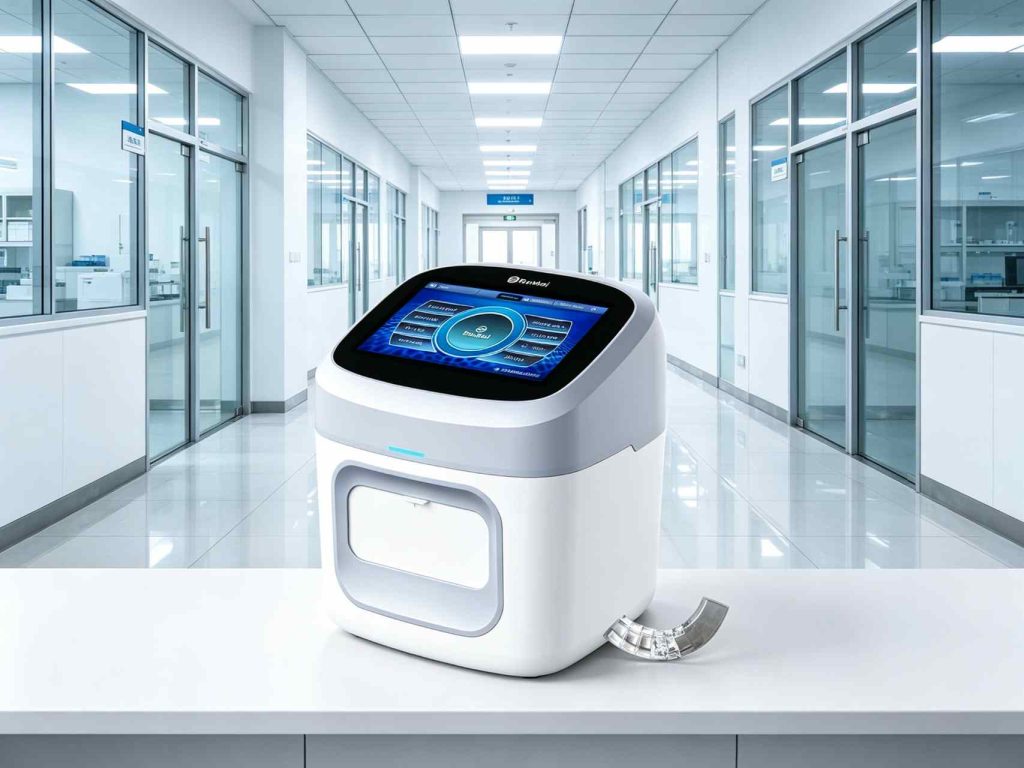
Looking for a reliable option? The 5-part differential hematology analyzer from RunMei Health is built for accurate, efficient blood analysis.
RDW and MCV Together: The Diagnostic Matrix
Combine RDW with MCV for a powerful first-pass screen: low MCV with high RDW suggests iron deficiency; low MCV with normal RDW suggests thalassaemia trait; high MCV with high RDW suggests B12/folate deficiency or haemolysis; normal MCV with high RDW suggests early deficiency or mixed anaemia. This two-variable pattern recognition beats relying on MCV alone and sharpens your differential before confirmatory tests.
Limitations and Caveats
RDW is not diagnostic on its own — it flags heterogeneity, not a specific disease. Recent transfusions, cold agglutinins, and very high reticulocyte counts all raise RDW. Always interpret it alongside MCV, the reticulocyte count, the film, and clinical context. Used right, it is one of the highest-value parameters your analyzer prints for free.
RDW Beyond the Anaemia Workup
RDW does more than decode anaemia. Elevated RDW is an independent predictor associated with cardiovascular events, adverse outcomes in heart failure, and higher inflammatory burden in some studies, and it rises with iron-poor states even when haemoglobin is still normal. Some clinicians track RDW alongside ferritin to catch early iron depletion in frequent blood donors or menstruating women before anaemia develops. It is a cheap, already-printed parameter with more clinical reach than its name suggests.
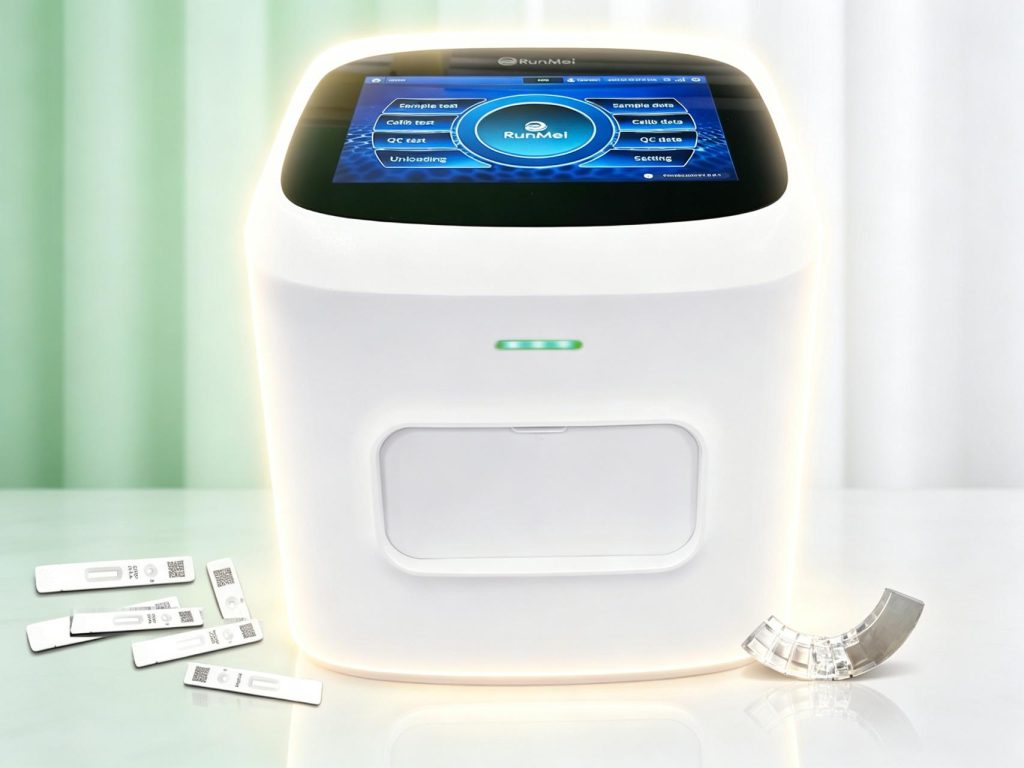
Looking for a reliable option? The portable multi-function blood analyzer from RunMei Health is built for accurate, efficient blood analysis.
Practical Reading in Your Workflow
Make RDW part of your standard interpretation checklist rather than a back-bench curiosity. Note it next to MCV, glance at the red cell histogram the analyzer prints, and look for the broadened curve that confirms anisocytosis. When RDW is high, the film is worth a look even if the patient is asymptomatic. Keep in mind that RDW varies naturally in infancy as cell sizes change, so use age-appropriate ranges and interpret patterns rather than reacting to an isolated surprise. Above all, never change treatment on RDW alone — it steers the investigation, but the film and confirmatory tests still carry the final say.
For a dependable solution, explore the 5-part veterinary blood analyzer from RunMei Health.
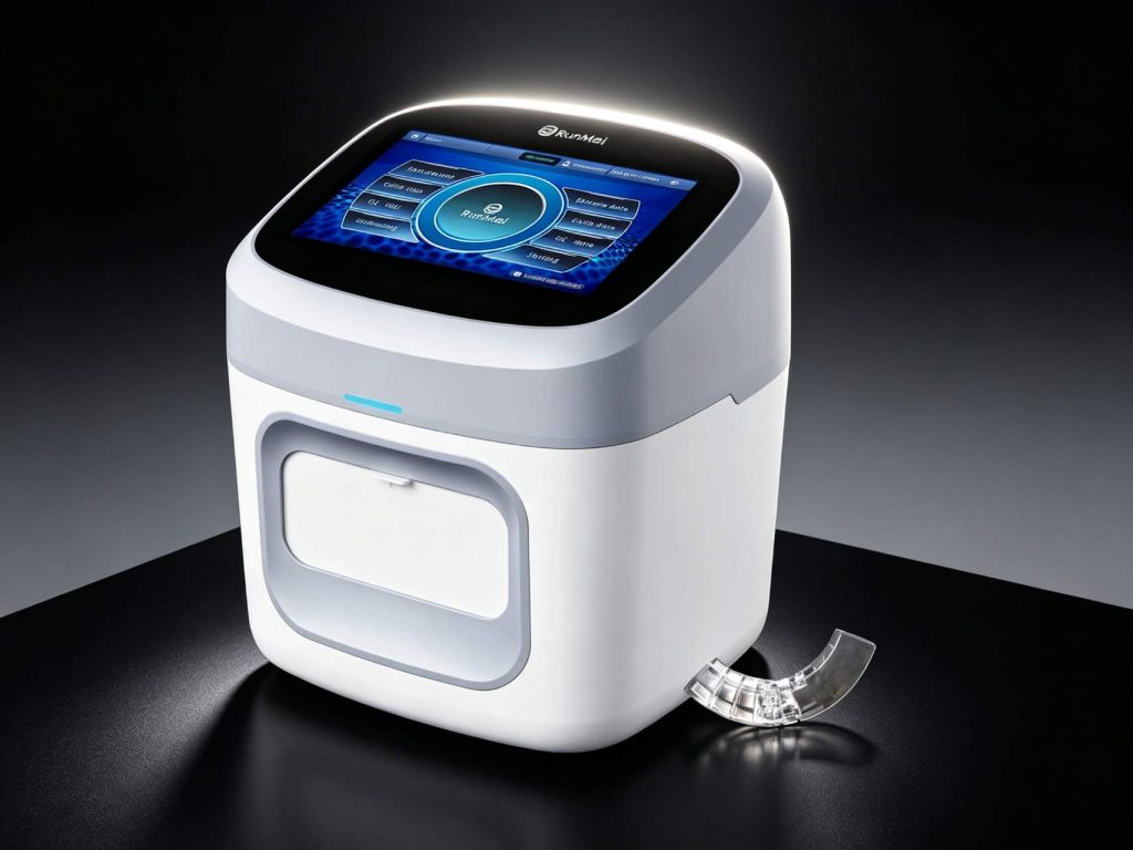
For a dependable solution, explore the RunMei diagnostic instruments from RunMei Health.
الأسئلة المتداولة
Q: What causes a high RDW?
A: A high RDW reflects unequal red cell sizes and appears in iron-deficiency anaemia, mixed deficiencies, haemolysis with reticulocytosis, recent transfusion or therapy, and myelodysplastic syndromes. It is a flag of cell-size heterogeneity that narrows the differential when read with MCV and the reticulocyte count.